
Anatomy Content Now Available in the Didactic (PA-1) Qbank


Last week we added new anatomy content to your Didactic Qbank subscription.
Did you see it?
Here’s an example from the new anatomy content:
Which structure does the left recurrent laryngeal nerve loop around?
A) Arch of the aorta
B) Left internal carotid artery
C) Left vertebral artery
D) Right subclavian artery
Answer A
Explanation: The vagus nerve (cranial nerve X) passes through the neck in the carotid sheath with the internal jugular vein and the common carotid artery. The recurrent laryngeal nerves are branches of the vagus nerve. The right recurrent laryngeal nerveloops around the subclavian artery, whereas the left recurrent laryngeal nerveloops around the arch of the aorta. The vagus nerve innervates the intrinsic muscles of the larynx, esophagus, and trachea and sends parasympathetic fibers to the heart. The sympathetic fibers to the head and neck begin in the thoracic region of the spinal cord and must ascend into the neck. The fibers ascend by entering into the cervical sympathetic chain, which has three ganglia (collections of nerve cell bodies): the superior, middle, and inferior cervical sympathetic ganglia. Sympathetic fibers synapse within these ganglia, and postsynaptic fibers continue into the head and neck, traveling closely along arteries. The superior cervical ganglion is located anterior to the C1–C4 cervical vertebrae and posterior to the internal carotid artery. The postganglionic nerves that originate from the superior cervical ganglion include the internal carotid nerve, external carotid nerve, nerve to the pharyngeal plexus, superior cardiac branch, and gray rami communicantes. The middle cervical ganglion is absent in some individuals but is located anterior to the inferior thyroid artery and the C6 vertebra when present. Its postsynaptic branches include gray rami communicantes, thyroid branches, and middle cardiac branch. The inferior cervical ganglion is anterior to C7. Its postsynaptic fibers include gray rami communicantes, branches to the vertebral and subclavian arteries, and the inferior cardiac nerve.
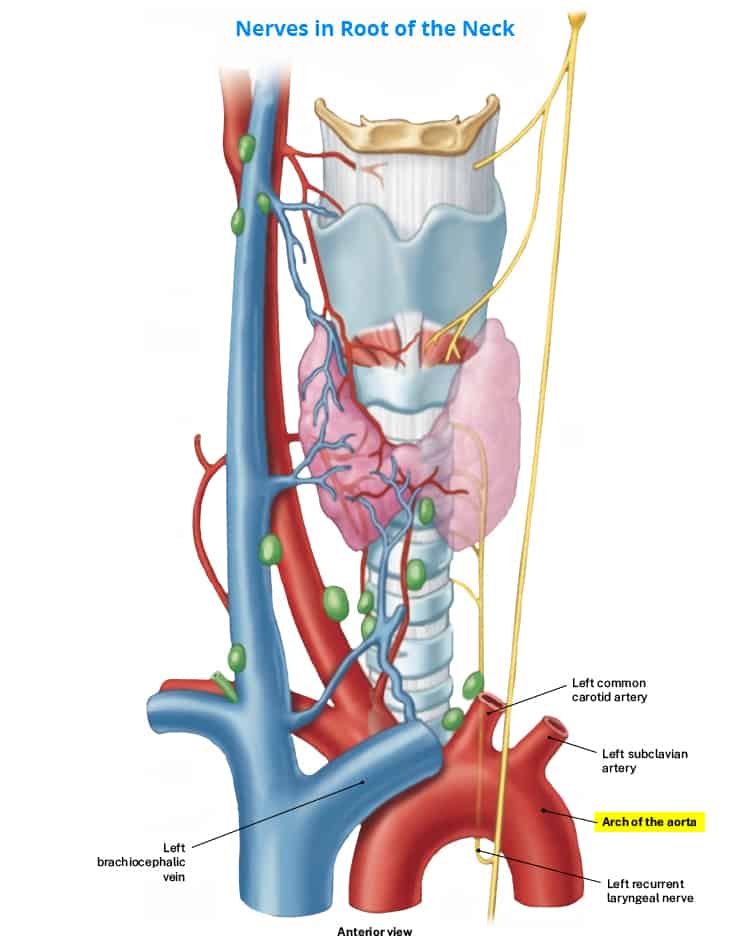
The left internal carotid artery (B) ascends the neck into the brain after the bifurcation of the common carotid artery. The postsynaptic sympathetic fibers after the superior cervical ganglion travel along the internal and external carotid arteries. Neither recurrent laryngeal nerve loops around the internal carotid artery. The left vertebral artery (C) is a branch of the left subclavian artery that supplies blood to the posterior part of the brain. The recurrent laryngeal nerves do not loop around the vertebral arteries. The right subclavian artery (D) supplies blood to the right upper extremity. The postsynaptic sympathetic fibers of the inferior cervical ganglion innervate the subclavian arteries. The right recurrent laryngeal nerve loops around the right subclavian artery, however, the left recurrent laryngeal nerve loops around the arch of the aorta.
One Step Further
Q: Which arteries do the superior cervical ganglion postsynaptic fibers travel with?
A: Internal and external carotid arteries.
Reference
Moore KL, Agur AMR, Dalley AF. Neck. In: Essential Clinical Anatomy. 5th ed. Lippincott Williams & Wilkins; 2015:581–626.
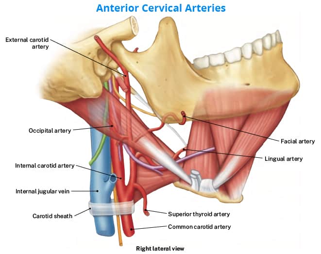
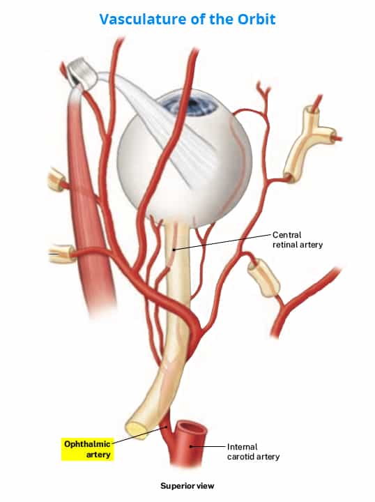
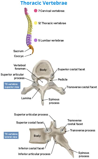
Here’s a sneak peek into other images from the anatomy content:

Current subscribers to Rosh Review’s Didactic (PA-1) Qbank receive this content for free!

Comments (0)