Introducing the Newest Qbank for the ABEM Advanced EM Ultrasonography Examination

As of June 1, 2021, ABEM has opened applications for the new Focused Practice Designation (FPD) in Advanced EM. A trusted option for those preparing for the Advanced EM Ultrasonography (AEMUS) Examination now exists: the Advanced Ultrasound Qbank, designed by three leaders and advocates in emergency medicine.
The Qbank is the vision of Dr. Resa E. Lewiss, Dr. Amy Zeidan, and Dr. Penny Lema after realizing the need for a high-quality resource for ABEM-certified physicians and ultrasound fellows studying for the AEMUS FPD exam, first offered in March 2022. Beyond this need, they recognized the importance of highlighting authors with unique journeys, expertise, and knowledge. Using the Advanced Ultrasound Qbank is not only a way to easily prepare for the FPD exam but also an opportunity to support the work of this community.
Why create an Advanced Ultrasound Qbank?
Drs. Lewiss, Zeidan, and Lema realized the need for a resource that prepared ultrasound fellows and faculty for their AEMUS FPD exam and highlighted the diversity of expertise in the ultrasound community. They are inspired by and pay homage to the legacy of the first three emergency ultrasound fellows: Beth Thomas, MD, Verena Valley, MD, and Mary Beth Phelan, MD. Based on this shared mission, the three creators brought their vision to Rosh Review to create the Advanced Ultrasound Qbank.
The Advanced Ultrasound Qbank is the first Qbank geared toward advanced ultrasound topics to prepare emergency ultrasound fellows for the AEMUS FPD examination.
Who is the Advanced Ultrasound Qbank for?
The Qbank was created for ABEM-certified physicians and ultrasound fellows taking the AEMUS FPD exam. However, students, residents, and clinicians of all levels can use it to build advanced ultrasound knowledge.
What does the Advanced Ultrasound Qbank contain?
The Qbank includes peer-reviewed questions and explanations using real case scenarios with accompanying ultrasound images and videos. This Qbank can help you solidify your advanced ultrasound knowledge about what normal examinations look like versus abnormal findings that could compromise a patient’s health.
Which topics are included in the Advanced Ultrasound Qbank?
The Qbank questions are aligned with the ABEM distribution of questions for the FPD examination. The FPD will cover the core content of advanced emergency medicine ultrasonography.
| Categories | Distribution |
|---|---|
| Physics and Technical Aspects of Ultrasonography | 10 ± 5% |
| Anatomic/Diagnostic Ultrasound | 60 ± 5% |
| Procedural Ultrasound | 10 ± 5% |
| Clinical Ultrasonography Training with Non-Emergency Medicine Specialties* | 0% |
| Education and Research Skills | 10 ± 5% |
| Administration and Quality | 10 ± 5% |
How are the Qbank questions structured?
Practice questions are written to resemble the format and topics on your exam. All Rosh Review Qbanks include detailed explanations for the correct and incorrect answer choices with teaching images (and videos), hyperlinked references, and performance analytics.
Example:

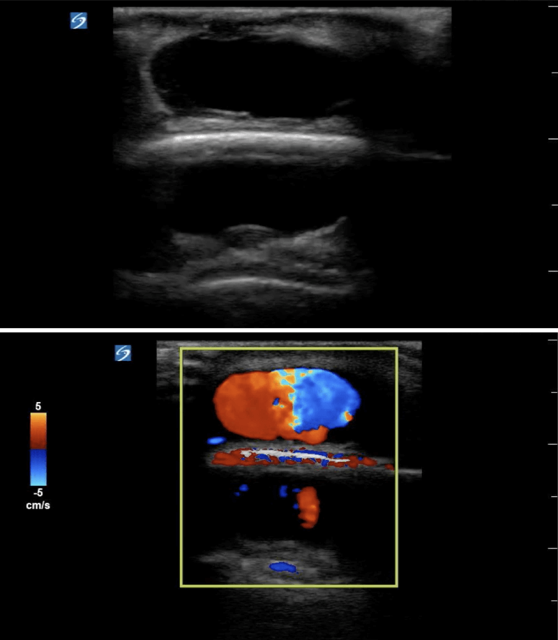
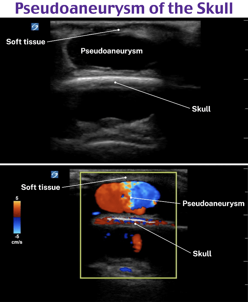
A 60-year-old man presents to the emergency department with a painful lump to the back of his head. 2 weeks prior he was hit in the head with a baseball bat and recently got his sutures removed. On physical exam he has a tender and fluctuant mass. The skin exam is limited because of his hair. A point-of-care ultrasound is performed as shown above. What is the next best step?
A) Antibiotics and follow-up with primary care doctor
B) Needle aspiration
C) Perform an incision and drainage and arrange for follow-up
D) Perform color Doppler of the mass prior to any intervention
Answer: D
Soft tissue ultrasound is performed using a high-frequency linear transducer. In this grayscale image, the mass appears as an anechoic rounded structure making it indistinct from a cyst, seroma, hematoma, abscess, or vascular anomaly. The mass is also on top of the scalp and the hyperechoic stripe of the scalp creates a mirror artifact. Performing color Doppler on any mass will identify if there is a source of blood flow within the cavity. Color Doppler can also help delineate a lymph node from other pathology. The image above is a pseudoaneurysm that formed after trauma. Pseudoaneurysms occur due to extravasation of blood outside of the vessel wall and into the perivascular space.

Antibiotics and follow-up (A) as well as incision and drainage (C) are incorrect answers because this is a pseudoaneurysm. An abscess is also an anechoic or hypoechoic fluid collection within a rounded cavity and often has irregular borders. Without using color Doppler, it can be indistinguishable from other fluid filled masses.
Needle aspiration (B) is also an incorrect answer because puncturing the pseudoaneurysm may cause more complications including external hemorrhage. There are a few ways to treat a pseudoaneurysm. First compress the mass using a linear transducer for approximately 20 minutes until pseudoaneurysm communication is closed. Another treatment option is injecting thrombin into the pseudoaneurysm. If the first two treatments fail then the pseudoaneurysm may require surgical intervention.
Where can I get started?
You can learn more about the Advanced Ultrasound Qbank here.


Comments (0)