
Sports Medicine Module For the ABFM Certification Exam Is Now Ready


Introducing the new ABFM Sports Medicine Content-Specific Module, available to Family Medicine residents, residency programs, and practicing physicians. This module is best suited for Family Medicine physicians looking for a focused, high-yield Qbank review for ABFMs Content-Specific Modules required during the certification exam. This content is novel, and not included in Rosh Review’s main Family Medicine Qbank or mock exams.
Family Medicine programs that subscribe to Rosh Review also gain access to the Content-Specific Modules in their PD Dash.
Categories include:
- Pre-participation evaluation
- Common conditions in athletes
- Acute injuries in athletes
- Overuse injuries in athletes
Content is organized by task:
- History and Physical
- Diagnostic studies
- Diagnosis
- Health maintenance
- Clinical intervention
- Clinical therapeutics
- Scientific concepts
Here is an example:
A 6-year-old boy is being evaluated for elbow pain following a fall. He was playing basketball and fell on an outstretched arm. Physical examination of the right elbow shows swelling, pain and tenderness, and limited range of motion. A right radial pulse is palpable. Which of the following would most likely confirm the diagnosis of this patient’s condition?
A. Anteroposterior, lateral, and oblique radiographs
B. Arthrography
C. Magnetic resonance imaging
D. Ultrasound with Doppler flow
Answer A
Falling on an outstretched arm, resulting in pain and limited range of motion, is strongly suggestive of a supracondylar fracture. Supracondylar fractures are the most common pediatric elbow fracture (most commonly between the ages of 5 and 10 years). In children, the supracondylar region encompasses an area of thin, weak bone located in the distal posterior humerus, which is susceptible to traumatic injuries. Radiographs should be obtained in children with suspected supracondylar fracture. Condylar fractures typically require 4 views (anteroposterior, lateral, and obliques) to be fully characterized. Analgesia should be administered prior to obtaining radiographs and caution should be taken to avoid excessive manipulation of the affected extremity.
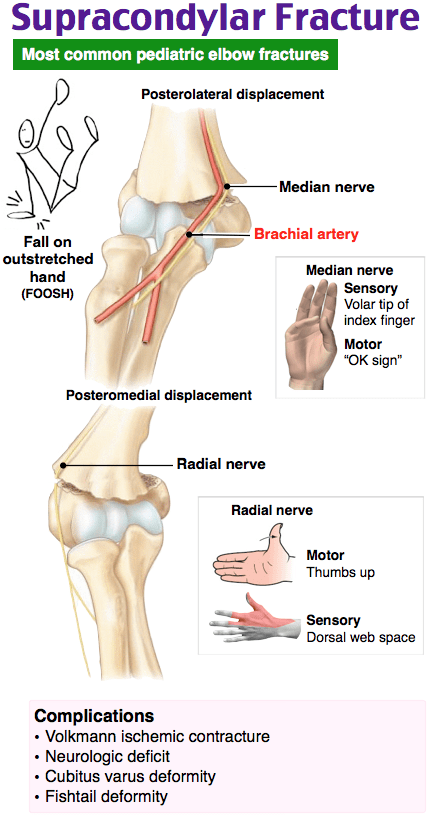
Arthrography (B) and magnetic resonance imaging (C) may be needed to differentiate lateral condylar from transphyseal fractures. However, they are not used to confirm the diagnosis of a supracondylar fracture. An ultrasound with Doppler flow (D) would be beneficial if there was evidence of vascular compromise (e.g., decreased or absent radial pulse).
One Step Further question:
What is a potentially feared complication of a supracondylar fracture?
Answer:
Compartment syndrome.
Since these categories and tasks are integrated into our improved performance and feedback page, you have access to robust data to help you fine-tune your studying and resources to help increase your Family Medicine Certification Exam score by 100 points.
Or visit the main Family Medicine Qbank page to learn about our ABFM Certification Qbank
Study on…
The Rosh Review Team
A bolus of confidence. A lifetime of knowledge.

Comments (0)