A is the correct answer. Why?
Get More POCUS Questions
Start today with a 10-day free trial.
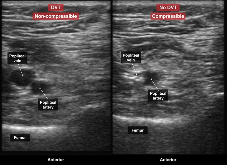
A A noncompressible segment in the popliteal vein with visible intraluminal echoes
B An anechoic lumen in the popliteal vein with full compressibility
C Compressibility of the common femoral vein with no visible thrombus
D Hyperechoic material within the superficial femoral vein with partial compressibility
Explanation
The hallmark finding on ultrasound for deep vein thrombosis is a noncompressible vein segment, which is indicative of a thrombus obstructing the venous lumen. The presence of visible intraluminal echoes further supports the diagnosis, as they represent the thrombus itself within the vein. In this case, the popliteal vein is the location of interest due to the patient’s calf symptoms, and the inability to compress this vein segment on ultrasound is the most specific finding for DVT. This imaging characteristic is crucial for the diagnosis and guides the subsequent management, which typically includes anticoagulation therapy to prevent clot propagation and embolization.
An anechoic lumen in the popliteal vein with full compressibility (B) indicates a patent vein and essentially rules out the presence of DVT at that location. This option might be erroneously selected by those who confuse the ultrasound appearance of a normal vein with that of a thrombosed vein.
Compressibility of the common femoral vein with no visible thrombus (C) is suggestive of a patent vein without evidence of DVT. In the setting of DVT, one would expect to find a segment of the vein that is not compressible due to the presence of a thrombus.
Hyperechoic material within the superficial femoral vein with partial compressibility (D) may indicate a chronic or partially recanalized thrombus rather than an acute DVT. While this finding warrants further investigation, it is not as diagnostic of acute DVT as a completely noncompressible vein with visible thrombus.